Quicklinks
Interested in using the CIC? Prerequisite: Training is required for all investigators prior to first time usage of any specific equipment in our facility.
Please fill out a Service Request Form (see above).
Available Instruments
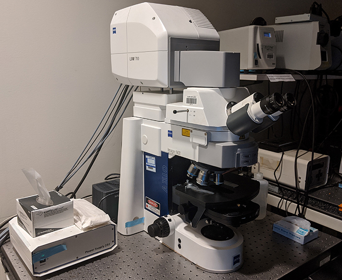
Zeiss 700 laser scanning confocal microscope
This upright confocal microscope is suitable for acquiring higher resolution images. It is equipped with a FRAP module.
Lines: 405, 488, 555, 635nm
Objectives:
Air: 10x (0.3NA)
Oil: 25x (0.8NA) multi-immersion and 63x (1.4NA) oil.
Located in CLS 13035
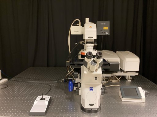
Zeiss LSM 980 with Airyscan 2 laser scanning confocal microscope
The Zeiss LSM980 is a laser scanning confocal microscope, enabling full 3D imaging of both live and fixed samples. The airyscan detector enables faster acquisition and higher resolution than typical confocal.
- Airyscan detector provides near super-resolution (120nm)
- Rapid z-stack acquisition (via a piezo)
- Motorized stage for tilescanning and mosaic imaging
- 6-channel simultaneous acquisition
- 6 laser lines (405, 445, 488, 561, 594, and 639 nm)
- Objectives: 10x/0.3 air, 20x/0.8 air, 40x/1.1 WI, 63x/1.4 Oil
- Multiplexing for acquisition rates of up to 47.5 fps
- Stage-top incubator for live-cell imaging
- Spectral unmixing
Please cite NIH grant #S10OD030322 if using this instrument for a publication. Thank you!
Located in CLS 13035
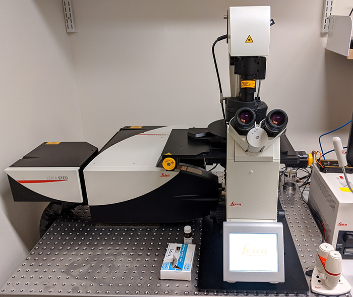
Leica TCS-SP8 confocal & STED super-resolution microscope
This system is a super resolution microscope that can achieve resolutions of 50nm. It has 5 detectors, 3 of which are high-sensitivity hybrid (HyD) detectors. It is equipped with an OkoLabs incubation chamber for live-cell imaging.
Lines: White light laser tunable 470-670nm, Argon laser
Objectives:
Air: 10x, 20x
Oil: 60x, 100x
Located in CLS 13035
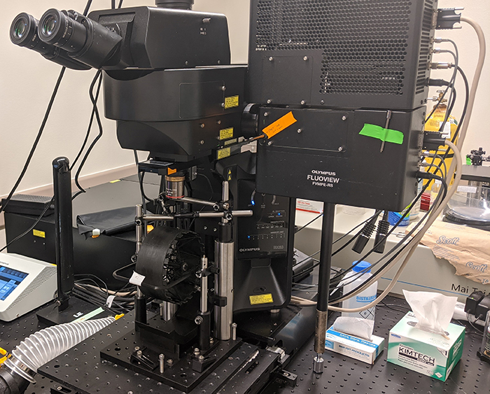
Olympus MPE-RS multiphoton microscope
This upright microscope is designed for in-vivo live imaging of anesthetized or awake-behaving mice and is equipped with a MaiTai tunable IR laser (640-1040nm), 2 high-sensitivity GaAsP detectors (green & red channels), and a Hamamatsu camera for epifluorescence imaging. Custom rigs are available.
Available equipment: nPoint Z Piezo adapter (for high speed volumetric imaging).
Objectives: 4x (0.28NA) air, 10x (0.5NA) water, 20x (0.5NA) water, 40x (0.5NA) water, & 25x (1.0NA) water objective.
Objectives:
Air: 4x (0.28NA)
Water: 10x (0.5NA), 20x (0.5NA), 40x (0.5NA), & 25x (1.0NA)
Located in Enders SB68
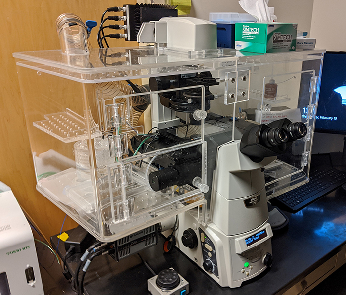
Nikon Ti-Eclipse inverted microscope
This fluorescence inverted microscope is equipped with a humidity-controlled box and an incubator unit that makes it ideal for long-term imaging of living samples. It is equipped with a motorized stage for tile-scanning & high quality camera (Andor Zyla sCMOS camera).
Imaging modes: epifluorescence (DAPI, GFP, RFP, and Cy5 filter sets), brightfield, phase and differential interference contrast (DIC).
Lines: 405, 488, 555, 647nm
Objectives:
Air: 1x (0.04NA), 10x (0.3NA), 20x (0.5NA), and 40x (0.75NA)
Multi-Immersion: 20x (0.75NA, multi)
Oil: 40x (1.3 NA), 60x (1.4NA), and 100x (1.3NA)
Located in CLS 13035
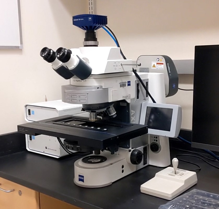
Zeiss Axio Imager.Z2 Fluorescence Microscope
The Axio Imager.Z2 is an upright widefield microscope that offers LED epifluorescence (385/430/475/555/630/735nm) and halogen brightfield light sources, highly sensitive AxioCam camera and a motorized stage that can hold up to 8 standard slides concurrently for high-throughput imaging as a slide scanner.
Filters: DIC/TL, DAPI, GFP, AF568/TexasRed, 90HE (DAPI/GFP/Cy3/Cy5), 110HE (DAPI/GFP/C3.5)
Objectives: 10x air, 40x oil, 63x oil, 100x oil.
Located in CLS 13035
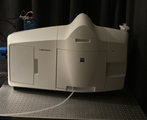
Zeiss Lightsheet 7 Microscope
The Zeiss LS7 is lightsheet microscope, enabling full volumetric imaging of transparent samples, both live and fixed. Lightsheet requires very particular sample preparation – contact core staff for consultation.
- 2-channel simultaneous acquisition
- Laser lines: 405, 488, 561, 638 nm
- Illumination optics: 5x and 10x
- Acquisition objectives: 10x and 20x water objectives for live imaging. 2.5x, 5x,and two 20x/1.0 objectives (RI of 1.45 and RI of 1.53) for cleared samples
- Acquisition rate: up to 57 fps
- Internal incubator for live-cell imaging
- Offline PC for data reconstruction
Located in CLS 13035
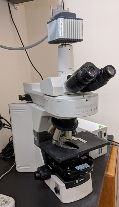
Nikon 80i fluorescence microscope
This microscope offers fluorescence and epifluorescence light sources and is equipped with air objectives & a Hamamatsu CCD camera.
Imaging modes: epifluorescence (DAPI, fluorescein, GFP, rhodamine, Texas red, Cy3, and Cy5 filter sets), brightfield, darkfield, phase and differential interference contrast (DIC).
Objectives:
Air: 1x (0.04NA), 2x (.1NA), 4x (.02NA), 10x (0.3NA), 20x (0.5NA), and 40x (0.75NA) and 60x (.95NA)
Located in CLS 13035
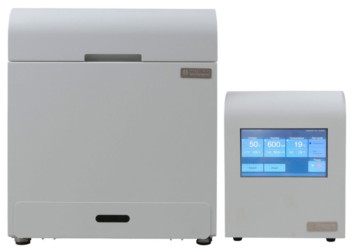
Life Canvas SmartClear clarity tissue clearing system
This system allows for rapid clearing of whole tissues through an automated system using a modified Clarity method.
Located in CLS 12145
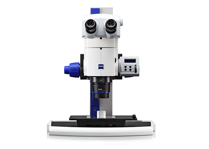
Zeiss SteREO Discovery Dissecting Microscope
Ideal for dissections, this microscope uses a variable LED transmitted light base for even illumination of specimens, supplemental fibre optic illuminators, and a rotatable nosepiece with a 0.3 x, 0.5 x and 1 x objectives with programmable motorized zoom. Video capable for protocol development.
Filters: UV, GFP, TexasRed
Located in CLS 12145
Workstation 1 (Imaris)
High-end desktop for processing graphics work running Imaris 9, FIJI (ImageJ), and Zen Lite. System can be used for 3D visualization, 3D rendering, and analysis of data.
Located in CLS 13035


